Original Source: https://www.fuzia.com/article_detail/767103/immunoblot-western-blot-an-effective-solution-for-protein
Western Blot is a process that people can use to determine the presence of proteins in cells and tissues. This test is often used in medical labs to detect if a sample contains cancer cells or abnormal tissue samples such as tumors. It is also used to measure the levels of different proteins in the blood. In addition, this technique made it possible for doctors to detect unknown viruses by looking at antibodies they produce against them through Western Blot.
What about Immunoblot?
Western Blot is a technique that uses antibodies to detect target proteins in cells and tissues. It is based on the principle that antibodies bind with the target protein. For example, during immunoblot, a protein sample could be subjected to gel electrophoresis, where it would be separated by size (see also Differential display). The resulting gel would then be transferred onto a membrane where the protein of interest would be transferred by electrostatic force (by applying voltage). Once there, tagged antibodies specific to the target protein can bind. These are usually tagged using radioactive isotopes such as Radioactive iodine or radio-iodinated amino acids. These isotopes emit radiation that the radiometer can detect.
Detection and Visualization:
One of the techniques used to detect bound antibodies is Autoradiography. This technique uses photographic film exposed to radiation from radioactive isotopes. However, this technique makes it impossible to visualization of antibodies that did not bind to the protein of interest. Thus, researchers like Kendrick Laboratories have devised other techniques that would also visualize non-bound antibodies. One such technique is Western blotting which involves transferring non-bound antibodies from a gel onto a membrane. Visualizing and identifying these non-bound antibodies would make it possible to examine all the proteins in a sample.
Why Choose Immunoblot Western Blot?
Immunoblot Western Blot is a type of optical Immuno detect used to identify and quantify the number of proteins. This technique uses antibodies designed to bind with a specific protein as it migrates in a gel. The proteins are transferred from the gel to the blotting membrane, where antibodies can selectively detect them. Western blotting is one of the most popular techniques used by scientists to inspect different proteins and proteins involved in disease episodes.
How Does Immunoblot Western Blot Work?
Western Blot would begin with one protein moving from the gel onto a membrane. Next, tagged antibodies have been added that target that certain protein for detection. The binding antibodies will be electrically charged and are brought close to the membrane using an electric current. Non-bound antibodies are then washed away with a buffer solution before visualization of the bound antibodies through Autoradiography.
Procedure:
- Get Electrophoresis:
One of the first steps when conducting a western blot would be to prepare a sample of cells or tissues. Next, the proteins in these samples are separated by electrophoresis.
- One-Step Western Transfer:
Next, the proteins from the gel should be transferred to the membrane in one step. For example, the proteins can be transferred by casting off access before electrotransfer to the membrane. The membrane should then be incubated by people with tagged antibodies specific to certain proteins that bind with it. These reaction products can then be detected and visualized through Autoradiography.
- Protein Staining:
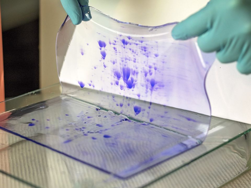
One of the ways that antibodies can be detected is by staining the membrane with a specific dye, such as Pierce’s Violet. The procedure for western blotting is easy to conduct. The steps are straightforward to follow with the help of instructions and labels provided with each kit. The proteins can be linked to antibodies, etc., using radio-iodinated nucleotides, which is one of the reasons western blotting is considered a powerful technique.
- Blocking:
Blocking the transfer of proteins from laboratory slides to the membranes can be done correctly because the membrane may contain antibodies binding with a target protein. However, if these antibodies are bound tightly with target proteins, it may hamper the transfer of targeted proteins in western blotting.
- Marking:
Some other steps need to be followed as part of western blotting. Clean-up should be part of this procedure. Moreover, markers should be added by people nto membranes to allow for easy visualization and identification of specific antibody bands.
- Incubation:
Antibodies tagged with radioactive isotopes should be added at the right time to allow for proper binding with target proteins. As a result, there should be proper monitoring of these reactions.
Other factors:
People should follow some other steps in washing solutions. First, these solutions should contain buffers that properly clean the membranes. Secondly, they should also contain other reagents, such as detergents that have protease, among other enzymes that can break down the antibodies into smaller polypeptides.
Conclusion:
Western blotting is one of the most powerful techniques people can use to test protein functions and search for biomedical applications. For example, the technique can detect different proteins and their interactions to understand disease mechanisms. If you want to learn about Immunoblot Western Blot, click here.

